Brow Lift
The supporting actor of upper-face feminization, not the hero. Most of what makes a forehead "read feminine" is the bone underneath — the supraorbital ridge, frontal bossing, and the angle from nose to forehead. The bone is reshaped by forehead contouring. A brow lift refines the soft-tissue position over that new bone.
This is why brow lift in FFS is almost never a standalone operation. In the largest published forehead-type series (Ousterhout, 1987), about 82% of FFS patients have a Type III forehead that requires bone work. That bone work needs an open incision. Once the forehead is already exposed, the brow lift is performed through the same access "for free" in incisional terms.
There's also a "relative lift" effect: when the supraorbital rim is shaved or set back, the brow ends up sitting higher relative to the new bone position even before any surgical suspension. Some patients with Type I or borderline foreheads achieve adequate brow elevation through bone reduction alone and need little or no brow lift on top.
Shape over height
The dominant feminine cue is the arched contour with the tail higher than the head — not raw millimeters of elevation. A flat brow lifted 6 mm straight up reads worse than a brow lifted 4 mm with a properly shaped lateral arch. The vector of pull and differential medial-vs-lateral fixation matter more than the average lift number.
The six approaches
Every brow lift answers three questions: how much lift, where's the scar, and what does it do to your hairline?
| Approach | Typical lift | Scar location | Hairline effect | Best FFS use case |
|---|---|---|---|---|
| Coronal (open) | 4–5+ mm, all positions | Behind hairline, ear-to-ear | Raises hairline | Type III forehead + already-low hairline |
| Pretrichial / trichophytic | 4–5 mm, all positions | At the hairline | Lowers hairline | Type III forehead + high or M-shaped hairline (≈85% of FFS patients) |
| Endoscopic | 1.6–3 mm | 3–5 small scars in scalp | Slight raise | Type I forehead, no hairline goals |
| Temporal / lateral | 2–3 mm, lateral only | Inside temple hair | Neutral | Lateral hooding only, or revision |
| Transblepharoplasty | ~2.5 mm, mostly lateral | In upper-eyelid crease | None | Modest lateral lift combined with blepharoplasty |
| Direct | Variable | Just above the brow | None | Generally avoided in FFS |
Coronal
Incision runs ear-to-ear several centimeters behind the frontal hairline. Maximum exposure for frontal sinus setback, supraorbital rim contouring, muscle modification (corrugator/procerus), and brow suspension all at once. Scar hidden in hair, but the technique inherently raises the hairline by the width of scalp removed — bad for patients who already have a high or recessed hairline. Sensory nerves are unavoidably divided behind the incision, producing a predictable patch of scalp numbness that often persists 9–12 months.
Pretrichial (trichophytic)
The workhorse approach for FFS. Incision placed at the hairline; the trichophytic version uses a 2–3 mm posterior bevel so hair grows through the scar. Closing the incision after scalp excision lowers the hairline at the same time. In a UCSF cohort of 46 trichophytic FFS procedures, mean brow elevation was 4.6 mm with simultaneous 5.9 mm of hairline lowering. About 85% of FFS patients are better candidates for this approach than coronal.
The honest trade-off: the scar is never invisible, even with the trichophytic bevel. Camouflage strategies include deferred hair transplantation (graft survival ~90% if delayed 3–6 months versus ~60% simultaneous) or makeup. Patients with progressive androgenic recession not yet stabilized by hormones / 5α-R inhibitors are poorer candidates, because the scar can become exposed if the hairline retreats. See also Hairline Advancement.
Endoscopic
Three to five small (1–2 cm) incisions behind the hairline; surgery done through a camera. Minimal scarring, less sensory loss, faster recovery — but typically only 1.6–3 mm of lift, and incompatible with significant frontal bone work. Fixation is achieved with bone tunnels, Endotine resorbable plates, sutures, or screws, with debated long-term durability.
Endoscopic brow lift use has declined at most high-volume FFS centers because the typical FFS patient needs Type III bone work that endoscopy can't accomplish. Reasonable for the minority with Type I foreheads, no hairline goals, and modest lift needs.
Temporal (lateral)
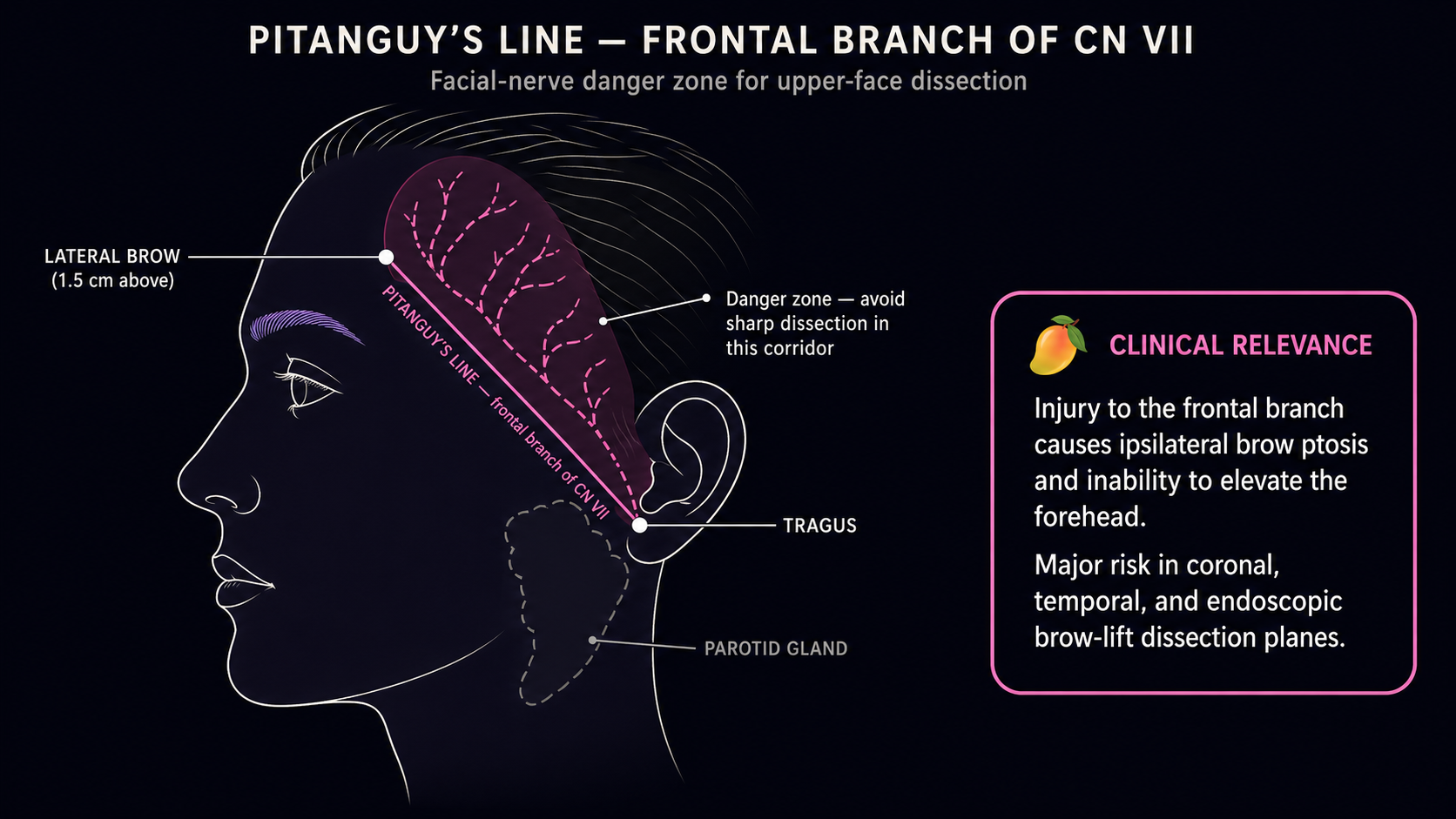
Small incisions hidden in temporal hair; elevates only the tail of the brow. Excellent for lateral hooding and creating a feminine arch when the medial brow is already adequate. Cannot lift the medial brow or address central forehead bone. Usually an adjunct or revision tool. Frontal branch facial nerve is the dominant injury risk — it runs along Pitanguy's line in this exact territory.
Transblepharoplasty (internal)
Sutures the lateral brow upward through the upper-eyelid blepharoplasty incision. Only ~2.5 mm of lift, mostly lateral. No scar visible in the forehead, no scalp numbness. Reasonable if you're already getting upper blepharoplasty and want modest lateral elevation, or for refining lateral brow position after primary FFS.
Direct (avoid)
Incision directly above the brow. Generally inappropriate in FFS for two reasons: trans women typically thin and shape brows during transition, removing the bushy hair that camouflaged the scar in the older male patients this technique was designed for; and the supraciliary skin is sebaceous and prone to widened, hypertrophic, hyperpigmented scars. One study reported 22.5% patient dissatisfaction with the scar. Reserve for asymmetric brow ptosis after facial nerve injury or salvage situations.
How much lift, and exactly where
The best published quantitative FFS data: UCSF 46-patient trichophytic cohort with mean elevation of 4.6 mm, essentially uniform across the medial canthus, midpupillary line, and lateral canthus. The medial brow remained slightly lower than lateral postoperatively, preserving a feminine arch. Commonly cited surgical range is 3–8 mm, with most operations at 4–6 mm. Surgeons typically over-correct by ~10–20% at the table because the brow drops modestly in the first 3 months as swelling resolves.
But raw millimeters are the wrong frame.
Classical (Westmore, 1974) target for the feminine brow:
- Medial head on a vertical line through the lateral nasal ala
- Tail along an oblique line from the ala through the lateral canthus
- Peak of the arch above the lateral limbus (outer edge of the iris)
Modern updates (including a 2018 fashion-model brow-position study) argue the peak has migrated more lateral, sitting between the lateral limbus and the lateral canthus, with the medial brow at or just above the rim rather than dramatically elevated.
The mechanism for creating that arch is the vector of pull, not the amount:
- Vertical vector → elevates medial as much as lateral → flat, "startled" or "Spock-brow" look
- Oblique superolateral vector (30–70° from horizontal) → preferentially elevates the tail → the feminine arch
Differential suturing or bone-channel fixation lets the surgeon dial in more lateral lift than medial. Common adjunctive maneuvers:
- Corrugator supercilii / procerus muscle weakening or resection — reduces the muscles that depress the medial brow and create the "11" glabellar lines
- Arcus marginalis release — selective release of the periosteum along the supraorbital rim
- Botox to corrugators/procerus — reinforces the surgical effect for 3–4 months; common adjunct
Some patients need no brow lift at all. Those with already-feminine brow positions, those whose Type III setback alone produces sufficient relative elevation, and the ~30% of people whose brows naturally elevate rather than ptose with age. A surgeon who proposes the same lift for every FFS patient regardless of starting anatomy is not individualizing.
Pre-existing asymmetry is the rule, not the exception — fewer than 2 mm of left-right brow difference is typically considered imperceptible. Good surgeons photograph and measure each side, plan asymmetric strip excision and differential fixation tension, and document expectations preoperatively.
Recovery
Recovery for a brow lift performed alongside forehead contouring is meaningfully longer than for an isolated cosmetic brow lift.
| Phase | What to expect |
|---|---|
| Days 0–5 | Swelling peaks. With Type III setback, periorbital swelling is dramatic — many patients' eyes swell shut for several days. Most surgeons prescribe a tapered oral steroid (Medrol dose pack). |
| Day 7–14 | Stitches/staples removed, often around day 7. Bruising transitions purple → green → yellow; gravity-tracks down to cheeks and even neck. |
| Weeks 1–2 | Significant swelling persists. Head-elevated sleep at 30–45° during this period. Most patients can conceal residual bruising with makeup by week two. |
| Weeks 2–6 | Heavy lifting (>10 lb / 5 kg) and strenuous exercise restricted; bending forward avoided for 2–3 weeks. Return to desk work: ~1 week (endoscopic), ~2 weeks (open brow lift alone), 2–5 weeks combined with full FFS. |
| Months 3–6 | Most external swelling resolved. Some brow drop from peak intraoperative position settles into the final result. |
| Month 6 | Final brow position typically apparent. |
| Months 9–12 | The conventional gold standard for assessing the bony FFS result, including final brow position. |
Numbness is the most under-appreciated part of recovery. With coronal or pretrichial approaches, sensory nerves are necessarily transected. A predictable patch of scalp numbness behind (coronal) or above (pretrichial) the incision is inevitable, often persists 9–12 months, and can be permanent in patches. Endoscopic approaches reduce but don't eliminate this; published rates ~2%. As nerves regenerate, itching is common and can be intense for weeks. A small subset develop persistent dysesthesias.
Scars mature over 6–12 months, sometimes up to 18, fading from pink toward final tone.
Risks and complications
In high-volume FFS centers, overall complication rates for forehead feminization (which includes the brow lift component) are low — 1.8% in a 168-case Spiegel series; 1.3% complications and 0.4% formal revision pooled across a 673-case systematic review. But "revision" is defined narrowly in those numbers; one large recent cohort reported up to 15% of patients had additional operations when scar revisions, hardware removals, and minor brow adjustments were counted.
| Risk | Reported incidence | Notes |
|---|---|---|
| Frontal branch facial nerve injury | ~0.1% endoscopic; up to ~0.7% transient, ~0.1% permanent in broader rhytidectomy data | Nerve runs along Pitanguy's line (0.5 cm below tragus to 1.5 cm above lateral brow); most vulnerable in temporal dissection. Most cases are neuropraxia, resolve over 3–6 months. |
| Sensory nerve issues (supraorbital, supratrochlear, deep branch of supraorbital) | Common; quantified rates vary widely | Usually resolve over 6–12 months; small permanent patches are common enough to be considered an expected trade-off, not a true complication. |
| Incisional alopecia | 2.2% coronal, 2.8% endoscopic, 1.5% temporal; up to 3% in one large FFS series | Strip of permanent hair loss along the scar. Trichophytic incision design and post-op transplantation are the standard mitigations. |
| Hairline displacement | Surgeon-choice-dependent | Coronal in a high-hairline patient = worse hairline; pretrichial in someone with progressing recession = exposed scar later. |
| Asymmetry / under-correction | Under-correction is the dominant reason patients return to OR per recent FFS revision data | One reason many surgeons prefer slight intentional over-correction at primary surgery. |
| Over-correction | Less common | Produces "surprised" look; rarely can pull eyelid skin upward enough to cause lagophthalmos (incomplete eye closure), usually temporary, more likely with simultaneous aggressive blepharoplasty. |
| Hematoma | ~1% in large series | Most managed nonsurgically. |
| Infection | Rare | Antibiotic-treatable in most cases. |
| Sinusitis (when combined with frontal sinus setback) | ~1% | See Forehead Contouring for full sinus complication discussion. |
| Hardware palpability / removal | ~5% palpable in one series; removal rare | |
| Dural injury / CSF leak / osteomyelitis | Very rare | Concentrated in Type III setback rather than the brow lift itself. |
Longevity
Brow position naturally relaxes over time — no brow lift is permanent.
- Endoscopic shows meaningful lateral relapse; one study documented only 50% surgeon satisfaction at 2+ years despite 70% patient satisfaction.
- Coronal / pretrichial generally hold better — more skin removed, stronger fixation.
- 5–10 years of durable result is a realistic informal expectation, with significant variability based on technique, fixation, genetics, sun exposure, and smoking.
Among fixation methods, bone tunnels with permanent suture have the best published profile. Cortical screws have the highest complication rate. Fibrin glue alone shows significant relapse by 9 months.
What to ask in consult
Three questions cut through the marketing language:
| Question | What a good answer sounds like |
|---|---|
| What's my Ousterhout forehead type, and how does that determine my incision choice? | Surgeon shows you a CT, classifies your forehead, explains why coronal vs pretrichial is right for your specific hairline. Not "we'll figure it out in surgery." |
| How many millimeters of lift, what vector — and how will you create the lateral arch? | Specific medial-vs-lateral targets; differential fixation; arch peak between lateral limbus and lateral canthus. Not just "I'll lift your brows." |
| What fixation method, and what are the realistic long-term outcomes? | Bone tunnels, Endotine, screws, sutures, glue — these are not equivalent. Surgeon should know their own retention data. |
Sources
Anatomic targets and historical aesthetics
- Ousterhout DK. (1987) — Feminization of the Forehead: Contour Changing to Improve Female Aesthetics (Plast Reconstr Surg) — original Type I/II/III classification; ~82% Type III prevalence.
- Westmore M. (1974) — Facial cosmetics in conjunction with surgery. Presented at the Aesthetic Plastic Surgical Society meeting, Vancouver, BC; May 7, 1974. (Conference paper, not formally indexed; classical feminine brow-target rule of thumb: apex above lateral limbus, medial and lateral ends level.)
- Jabbour S et al. (2018) — The Evolution of Eyebrow Morphology: The Westmore Model Revisited (Int J Dermatol) — fashion-model update arguing the modern feminine brow peak sits between lateral limbus and lateral canthus.
Operative series
- Garcia-Rodriguez L, Thain LM, Spiegel JH. (2020) — Scalp Advancement for Transgender Women: Closing the Gap (Laryngoscope) — UCSF 46-patient trichophytic FFS cohort; 4.6 mm mean brow elevation with simultaneous 5.9 mm hairline lowering.
- Spiegel JH. (2011) — Facial Determinants of Female Gender and Feminizing Forehead Cranioplasty (Laryngoscope) — 168-case Boston series; 1.8% overall complication rate.
- Khetpal S et al. (2024) — Feminization of the Forehead: A Scoping Literature Review and Cohort Study of Transfeminine Patients (Aesthetic Plast Surg) — 673-patient pooled literature: 1.3% complication rate, 0.4% formal revision.
Specific risks
- Ueda K, Harii K, Yamada A. (1994) — Long-term Follow-up Study of Browlift for Treatment of Facial Paralysis (Ann Plast Surg) — source of the 22.5% scar-dissatisfaction figure for direct brow lift; basis for avoiding the technique in FFS contexts.
Standards and frameworks
- Coleman et al. (2022) — WPATH Standards of Care v8 — facial gender surgery medical necessity criteria; hormones not prerequisite.