Penile-Inversion Vaginoplasty
The dominant primary technique for transfeminine genital reconstruction since Georges Burou's 1956 Casablanca clinic and Stanley Biber's adoption in the U.S. in the 1970s. The core mechanic has been stable for roughly seven decades: penile shaft skin is degloved, inverted, and used to line a surgically created perineal cavity; the glans (with its dorsal neurovascular pedicle preserved) is reshaped into a sensate neoclitoris; scrotal skin becomes the labia majora; and inner foreskin / preputial tissue becomes the clitoral hood and labia minora.
Modern series consistently report operative times of 4–6 hours, hospital stays of 4–7 days, canal depths of 10–15 cm with primary inversion alone, and orgasmic function in roughly 70–85% of patients with adequate follow-up. The dominant variable across surgeons is not the basic transposition map — that is largely standardized — but rather details of canal dissection plane, depth of glans burial, scrotal-flap geometry, urethral spatulation, and whether peritoneum or sigmoid is recruited when shaft skin is short.
This page covers the inversion technique itself. For peritoneal flap as primary or extension graft see Peritoneal Pull-Through. For minimal-depth construction see Vulvoplasty. For donor-site clearance see Pre-Op Hair Removal. For dilation and post-op care see Recovery & Dilation.
Primary inversion vs extension grafts
"Penile-inversion vaginoplasty" technically refers to the inversion mechanic, not the entire operation. When shaft skin is short, modern surgeons routinely add a full-thickness scrotal skin graft (FTSG) to the apex, a Davydov-style peritoneal flap, or — historically — sigmoid colon. These are extension techniques layered on top of inversion, not replacements for it. This page covers inversion as the primary mechanic; extension is covered in Peritoneal Pull-Through.
Donor-tissue mapping
The operation is in essence a re-mapping problem. Every recognizable structure of the post-op vulva is built from a specific piece of pre-op genital anatomy, with its blood supply (and for the clitoris, nerve supply) preserved. Understanding the map clarifies why pre-op hair removal of certain donor zones matters, why canal depth is bounded by donor skin available, and why revision labiaplasty is so commonly planned.
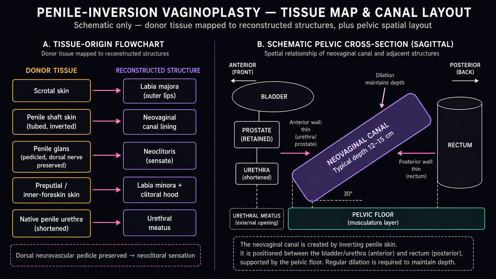
| Donor structure (pre-op) | Becomes (post-op) | Mechanic |
|---|---|---|
| Penile shaft skin (degloved as a tube) | Vaginal canal lining, anterior 2/3 to full length | Inverted (turned inside-out), apex sutured closed at the distal end, base anastomosed to the perineal opening; canal is packed with a stent against the new cavity |
| Scrotal skin (bilateral flaps, raphe excised) | Labia majora; in posterior-flap variants, the posterior canal wall | Hair-bearing scrotum is thinned of dartos and used as full-thickness flap; in Suporn-style techniques a tongue of scrotal skin lines the posterior introitus |
| Glans penis + dorsal neurovascular bundle | Neoclitoris | Wedge of glans (1–2 cm at widest) sculpted on a preserved dorsal NVB pedicle, tunneled to neoclitoral position, buried under preputial hood |
| Inner foreskin / preputial skin | Clitoral hood and labia minora | Preputial skin distal to the corona is preserved on the dorsal pedicle, draped over the buried neoclitoris, and split caudally to form labia minora |
| Penile (pendulous) urethra | Shortened urethral meatus; spatulated dorsal mucosa lines the anterior introitus | Spongiosum is largely resected to prevent post-op engorgement; urethra is shortened to perineal length, spatulated, and inset |
| Corpora cavernosa | Removed (not used) | Excised at the crura against the pubic ramus; proximal stumps oversewn — incomplete resection is the classic cause of post-op clitoral/introital bulging on arousal |
| Bulbospongiosus muscle and bulb | Mostly removed | Excised to flatten the perineum and prevent a "male" perineal contour or post-op pulsing at the introitus |
| Scrotal raphe / midline scrotal skin | Discarded or used as posterior commissure | The midline raphe is the densest hair zone and is typically excised; the resulting closure becomes the posterior fourchette |
The dorsal neurovascular pedicle is the rate-limiting step of the entire operation. The dorsal nerves of the penis (branches of the pudendal nerve) and the dorsal arteries run paired between Buck's fascia and the tunica albuginea on the dorsal aspect of the corpora. Preserving this bundle as a long pedicle while resecting the corpora beneath it is the defining technical maneuver of modern penile-inversion vaginoplasty (PIV); it is why the neoclitoris is sensate and why all reputable surgeons use loupe magnification or microsurgical dissection through this step.
The cavity and the canal
The neovaginal cavity is dissected sharply between the rectum posteriorly and the prostate / membranous urethra anteriorly, in the avascular plane behind Denonvilliers' fascia. The dissection is carried cephalad to the level of the peritoneal reflection, along the central tendon of the perineum and through the levator ani. Lateral boundaries are the levator and pelvic side walls.
The anterior wall is anatomically thin. The prostate and membranous urethra sit immediately anterior to it, separated from the canal only by Denonvilliers' fascia and the inverted skin graft. Three implications follow:
- Rectal injury is the most feared intra-operative complication. Reported in 1–4% of large series; Buncamper et al. (2016) reported 1.4% rectal injury across 475 cases at VU Amsterdam.
- Long-term dilation maintains anterior–posterior separation between canal and prostate / urethra. Canal stenosis can in rare cases progress to recto-neovaginal or urethro-neovaginal fistula.
- Prostate exams remain anatomically accessible per-neovagina post-op. The prostate is left in situ.
Canal depth is bounded by donor skin, not by surgeon ambition
Canal depth realistically achievable with primary inversion alone is 10–15 cm in most series. Numbers above 15 cm in inversion-only series should be read skeptically; they often refer to immediate intra-op stent depth, which contracts. The honest answer to "how deep will I be" is: a canal whose maximum is set by your stretched penile length plus available scrotum, typically 12–14 cm at one year of consistent dilation.
The four limiting factors:
- Donor skin length. Total useable shaft skin is set by stretched penile length. A 12 cm pre-op stretched shaft yields roughly that much skin tube; scrotal-flap extensions can add 3–6 cm.
- Body habitus. Android pelvis, deep perineum, and high BMI all reduce achievable depth — the dissection has to traverse more soft tissue before reaching the peritoneal reflection.
- Pre-op stretching. Some surgeons (historically the Suporn lineage) advise pre-op penile traction or weighted dilation. Evidence is anecdotal rather than RCT-grade.
- Whether a graft extension is added. FTSG to the apex, Davydov-style peritoneal flap, or sigmoid extend depth beyond what inversion alone yields; these are covered in Peritoneal Pull-Through.
Neoclitoris construction and sensation
The neoclitoris is built from a dorsal wedge of the glans (typically a triangular or shield-shaped segment, 1–2 cm at its widest) kept attached to the preserved dorsal neurovascular pedicle. The corpora are resected away from beneath the wedge; the wedge is rotated subcutaneously to the position of a clitoris (just inferior to the pubic symphysis, superior to the urethral meatus); preputial skin is draped over it as the hood.
Surgeon preference varies on how much of the glans is left visible. Some (Bowers, Schechter) emphasize a more visible glans tip; others in the Suporn lineage and Chettawut bury the neoclitoris more completely under the hood for a softer aesthetic. Both are defensible; neither has comparative outcome data favoring it.
Reported sensation and orgasm outcomes in cohort studies:
| Series | n | Setting | Orgasm capacity |
|---|---|---|---|
| Buncamper et al. 2015 | 49 | VU Amsterdam | 84% reported ability to orgasm; mean FSFI total 22.4 |
| LeBreton et al. 2017 | 169 | French cohort | ~78% orgasmic; sexual satisfaction high; lubrication low |
| Hess et al. 2014 | 119 | Essen series | 72% sexually active; of those, 78% reported orgasmic capacity |
| Lawrence 2005 | 232 | Multi-surgeon survey | ~85% reported ability to orgasm at least sometimes |
| Garcia et al. 2014 | small | Quantitative sensory | Light touch and pressure thresholds within cisgender clitoral baselines |
Long-term (>5 year) prospective sensation data are sparse; most series rely on cross-sectional surveys at variable follow-up. There is no high-quality data on whether neoclitoral sensation improves, plateaus, or declines beyond year 2. Lubrication is uniformly low in inversion-only series — inverted skin does not produce lubricant — and is one of the persistent reasons patients request peritoneal extension at primary or revision.
Urethral shortening and stream
The pendulous (penile) urethra is resected; the urethra is shortened to perineal length. Spongiosum is mostly excised to prevent it from acting as erectile tissue post-op — a retained spongiosum can produce a visible pulsing bulge or a "hood" of swelling at the introitus during arousal. The proximal urethra is spatulated (cut open longitudinally on its dorsal surface) and inset into the anterior introitus. In many techniques the spatulated urethral mucosa is used to line the anterior wet-tissue surface of the introitus, contributing wet-mucosal feel.
Stream issues are common in the first 3–12 months and usually resolve:
| Issue | Reported incidence | Course |
|---|---|---|
| Spraying / lateralized stream | 15–33% at 3 months | Most resolve by 6–12 months |
| Stream redirected downward | Typical post-op pattern | May require sitting slightly forward |
| Meatal stenosis | 4–14% at 3–9 months | Managed by dilation or revision meatoplasty |
| Persistent stenosis or fistula | <5% | Indication for urethral revision |
Buncamper (2016) and the Massie systematic review (2018) are the largest quantitative sources for these ranges.
One-stage vs two-stage protocols
Most North American and European programs perform PIV as functionally one and a half stages: a primary operation that creates the canal, neoclitoris, and rough labial form, plus a planned labiaplasty revision at 6–12 months to refine labia minora definition, clitoral hood shape, posterior fourchette, and any scar contractures.
Why revision is so commonly planned rather than treated as elective:
- After primary surgery, edema and post-op contraction distort labial position; surgeons cannot reliably build crisp labia minora and a defined hood at the same operation that creates the canal.
- Scrotal hair follicles that were incompletely cleared pre-operatively are easier to extract once tissue has settled.
- The introitus often needs minor relaxing or Z-plasty for entry comfort.
- It is the chance to address minor wound separation, granulation, or asymmetry from primary healing.
Variants:
- True one-stage (Suporn lineage, Chettawut, some European programs). All labial sculpting at the primary operation. Reduces total surgery count and travel burden; typically yields slightly less refined labia minora; less common in the U.S.
- Two-stage by design. Canal creation and labiaplasty deliberately separated by ≥6 months. Less common; sometimes used in patients with short shaft skin where stage 1 is canal lining and stage 2 is labial reconstruction with FTSGs.
'Single-stage' marketing language ≠ no revision
"Single-stage" in clinic copy typically means "primary surgery in one operation" — it does not promise no revision. Revision labiaplasty rates of 20–40% are typical even at high-volume centers (Gaither et al. 2018). Plan financially and logistically for a likely second operation; see Surgeons & Cost.
Major published technique variants
Citing by published series, not as endorsements. The wiki should not rank surgeons; the literature is what it is. These variants are convergent more than divergent — the basic transposition map is the same, and differences are in scrotal-flap geometry, depth of clitoral burial, single- vs two-stage labial sculpting, and how short shaft skin is handled.
| Surgeon / lineage | Location | Key published series | Distinguishing notes |
|---|---|---|---|
| Bowers | San Mateo, CA (formerly Trinidad — successor to Biber) | Bowers 2018 chapter, Principles of Gender-Specific Medicine 3rd ed. | Inversion with scrotal-flap depth extension; emphasis on visible clitoral architecture and labia minora at primary stage |
| Suporn / Chettawut | Chonburi / Bangkok, Thailand | Wangjiraniran et al. 2015 (Suporn 2004 conference series; Chettawut clinical reports) | Hybrid penile-inversion + large scrotal posterior-wall flap; long-tradition single-stage protocol; pre-op penile traction historically advised |
| McGinn / Rumer / Papillon | Pennsylvania | Rumer et al. 2015–2018 (cohort papers) | Mid-Atlantic high-volume programs; standard inversion with two-stage labiaplasty |
| Schechter | Chicago, IL | Schechter & Schechter 2016, Clin Plast Surg; Schechter 2016 textbook | Detailed published technique chapters; standard inversion with NVB-pedicled neoclitoris; advocates routine 6-month labiaplasty revision |
| Meltzer | Scottsdale, AZ | Book chapters; large clinical volume | One of the highest-volume U.S. PIV practices since the 1990s; standard inversion with scrotal-flap extension when needed |
| VU Amsterdam (Buncamper / Bouman / van der Sluis) | Amsterdam, Netherlands | Buncamper et al. 2016, Plast Reconstr Surg (475 cases); van der Sluis 2017 | Largest single-center European cohort; well-published complication rates (rectal injury 1.4%, neovaginal stenosis ~5%, fistula <2%); major source of peritoneal-extension data |
| Belgrade (Djordjevic et al.) | Belgrade, Serbia | Djordjevic et al. 2011, J Sex Med | High-volume European program with published outcomes on both PIV and sigmoid |
| Crane Center | Austin / San Francisco | Berli et al. 2017, JAMA Surg (review); Crane cohort papers | High-volume U.S. program; standard inversion plus published peritoneal-extension data |
For up-to-date pricing, travel logistics, and insurance posture, see Surgeons & Cost.
Operative time and hospital stay
- Operative time: typically 4–6 hours for primary PIV at experienced centers; 6–8 hours when peritoneal or sigmoid extension is added, when revision components are bundled, or in patients with short shaft skin requiring grafting.
- Hospital stay: typically 4–7 days in U.S. and European programs, with the canal packed and a urinary catheter in for the duration of the inpatient stay. Some programs (Mount Sinai, ambulatory European centers) discharge as early as day 3; Thai programs traditionally keep patients at affiliated recovery housing for 7–14 days.
- Catheter: 5–10 days; some surgeons leave a suprapubic catheter longer.
- Initial dilation: typically begun day 5–7, in hospital, with structured teaching before discharge — see Recovery & Dilation.
When peritoneum is added to inversion
Modern practice increasingly uses peritoneum as an adjunct to inversion rather than as a standalone primary technique — a Davydov-style peritoneal flap is pulled down to line the apex / anterior of the canal when shaft skin is insufficient. Most common in:
- Patients on long-term puberty blockers from Tanner stage 2 with limited natal genital tissue.
- Patients with a short stretched penile length (<10 cm) where inversion alone would yield <8 cm depth.
- Revision cases where the canal has stenosed and donor skin is exhausted.
Adding peritoneum is a distinctly bigger operation (laparoscopic or robotic abdominal access, longer OR time, peritoneal complications added to perineal complications) and is covered in Peritoneal Pull-Through. For the complication profile of canal stenosis, recto-neovaginal fistula, and revision indications, see Complications & Revision.
Where the literature is thin
- Long-term (>5 year) sensation, lubrication, and orgasmic function are under-studied. Most published outcomes are at 6–24 months.
- Direct comparative trials between surgeons or technique variants do not meaningfully exist; what exist are single-center retrospectives.
- Patient-reported outcome measures specific to gender-affirming genital surgery are still being validated (GENDER-Q in development).
- Pelvic-floor function, urinary continence, and sexual function in PIV patients 20+ years post-op are almost entirely uncharacterized.
- Mental-health and quality-of-life outcomes are well-published in aggregate but rarely linked to specific technique details.
A number of the most-quoted depth and orgasm numbers in patient-facing material trace back to single retrospective series with self-reported outcomes. This page marks numerical claims with cohort size and follow-up window where possible, and distinguishes primary published series from clinic marketing pages, which tend to round upward.
Sources
Primary cohort and technique papers
- Buncamper ME, van der Sluis WB, de Vries M, et al. Penile Inversion Vaginoplasty With or Without Additional Full-Thickness Skin Graft: To Graft or Not to Graft? Plast Reconstr Surg. 2017;139(3):649e–656e.
- Buncamper ME, Honselaar JS, Bouman MB, et al. Aesthetic and Functional Outcomes of Neovaginoplasty Using Penile Skin in Male-to-Female Transsexuals. J Sex Med. 2015;12(7):1626–34.
- van der Sluis WB, Bouman MB, de Boer NK, et al. Long-Term Follow-Up of Transgender Women After Secondary Intestinal Vaginoplasty. J Sex Med. 2016;13(4):702–710.
- Hess J, Rossi Neto R, Panic L, Rübben H, Senf W. Satisfaction with male-to-female gender reassignment surgery. Dtsch Arztebl Int. 2014;111(47):795–801.
- Massie JP, Morrison SD, Maasdam JV, Satterwhite T. Predictors of Patient Satisfaction and Postoperative Complications in Penile Inversion Vaginoplasty. Plast Reconstr Surg. 2018;141(6):911e–921e.
- Schechter LS, Schechter RB. Surgical management of the transgender patient: an overview. Clin Plast Surg. 2016;43(2):243–56.
- Schechter LS. Surgical Management of the Transgender Patient. Elsevier, 2016. Textbook chapters on PIV technique.
- Horbach SE, Bouman MB, Smit JM, et al. Outcome of Vaginoplasty in Male-to-Female Transgenders: A Systematic Review of Surgical Techniques. J Sex Med. 2015;12(6):1499–1512.
- Manrique OJ, Adabi K, Martinez-Jorge J, et al. Complications and Patient-Reported Outcomes in Male-to-Female Vaginoplasty: Where We Are Today. Ann Plast Surg. 2018;80(6):684–691.
Sensation and sexual-function studies
- Lawrence AA. Sexuality before and after male-to-female sex reassignment surgery. Arch Sex Behav. 2005;34(2):147–66.
- LeBreton M, Courtois F, Journel NM, et al. Genital Sensory Detection Thresholds and Patient Satisfaction With Vaginoplasty in Male-to-Female Transgender Women. J Sex Med. 2017;14(2):274–281.
- Garcia MM, Christopher NA, De Luca F, Spilotros M, Ralph DJ. Overall satisfaction, sexual function, and the durability of neoclitoral sensation following staged male-to-female genital reassignment surgery. Transl Androl Urol. 2014;3(2):156–62.
Complication and revision data
- Gaither TW, Awad MA, Osterberg EC, et al. Postoperative Complications following Primary Penile Inversion Vaginoplasty among 330 Male-to-Female Transgender Patients. J Urol. 2018;199(3):760–765.
- Wangjiraniran B, Selvaggi G, Chokrungvaranont P, et al. Male-to-female vaginoplasty: Preecha technique experience with 129 cases. J Med Assoc Thai. 2015;98(1):110–8.
- Djordjevic ML, Bizic M, Stanojevic D, et al. Urethral lengthening in metoidioplasty / vaginoplasty surgical technique reports. J Sex Med. 2011.
Reviews and historical foundation
- Bowers ML. Vaginoplasty in male-to-female transgender patients. In: Principles of Gender-Specific Medicine. 3rd ed. 2018.
- Berli JU, Knudson G, Fraser L, et al. What Surgeons Need to Know About Gender Confirmation Surgery When Providing Care for Transgender Individuals: A Review. JAMA Surg. 2017;152(4):394–400.
- Selvaggi G, Bellringer J. Gender reassignment surgery: an overview. Nat Rev Urol. 2011;8(5):274–82.
- Karim RB, Hage JJ, Mulder JW. Neovaginoplasty in male transsexuals: review of surgical techniques and recommendations regarding eligibility. Ann Plast Surg. 1996;37(6):669–75.
- Perovic SV, Stanojevic DS, Djordjevic ML. Vaginoplasty in male transsexuals using penile skin and a urethral flap. BJU Int. 2000;86(7):843–50.
- Jacoby A, Maliha S, Granieri MA, et al. Robotic Davydov Peritoneal Flap Vaginoplasty for Augmentation of Vaginal Depth in Penile Inversion Vaginoplasty. J Urol. 2019;201(6):1171–1176.
Standard textbook references
- Ettner R, Monstrey S, Coleman E, eds. Principles of Transgender Medicine and Surgery. 2nd ed. Routledge, 2016.
- Salgado CJ, Monstrey S, Djordjevic ML, eds. Gender Affirmation: Medical and Surgical Perspectives. Thieme, 2017.