Forehead Contouring
The single highest-impact bony procedure in FFS. The upper third of the face strongly drives perceived gender — brow bossing, the angle from nose-bridge to forehead (nasofrontal angle), and the apparent height of the hairline all read as masculine or feminine before any other feature does.
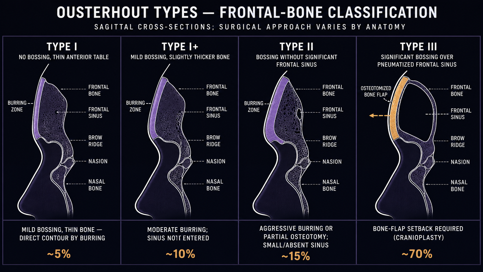
The taxonomy is Type I / II / III, named decades ago by Douglas Ousterhout. The number isn't a severity ranking — it's an anatomy classification. Which type your surgeon performs is determined by what's under your skin, specifically by your frontal sinus (a hollow air space behind the brow ridge), not by how feminine you want your forehead to look.
| Type | What's done | When it applies |
|---|---|---|
| Type I | Burring (grinding down) of the outer bone surface only | Frontal sinus is small/absent, or the bone over the sinus is thick enough to safely burr without breaking through |
| Type II | Limited burring of the bossing plus augmentation (filling in the dip above it with bone cement or implant material) | Brow projection is acceptable, but there's a flat or concave area above it. Niche; rarely used in modern practice. |
| Type III | The bone wall over the frontal sinus is cut free, reshaped, set back, and re-fixed with plates and screws | Sinus is large or its front wall is thin — burring alone would either be unsafe or insufficient. The most common modern operation. |
| Type I+ | Burring with focal full-thickness defects reconstructed using split bone graft | Newer hybrid: shallow sinus where most reduction can be done by burring, but a few small breakthroughs are expected and patched |
Type III is the modern workhorse
Most transfeminine patients have a sinus that's either large or has a thin front wall. In the largest contemporary single-center series (123 patients, 2024), 115 of 123 received Type III. Type II is now genuinely rare in modern outcome series. If a surgeon is recommending Type III for you, that's normal — it doesn't mean your case is severe.
How frontal sinus anatomy decides the operation
The frontal sinus is the variable. Two patients with identical-looking brow bossing on the outside can require different operations because their bone geometry differs:
- No sinus / very thick anterior table → Type I. The surgeon can burr bone away to your aesthetic endpoint without running out of safe material.
- Shallow sinus + thick table → Type I+. Most reduction is by burring, with focal patches where the burr breaks through.
- Large or thin-walled sinus → Type III. There isn't enough safe bone to burr; the anterior wall must be removed, reshaped, and set back.
CT-based studies show that masculine skulls average a more acute (sharper) nasofrontal angle and steeper forehead inclination than feminine skulls (mean 119.9° vs 133.5° in one study). The goal of contouring is to open that angle — but how much you can change it is constrained by the sinus anatomy underneath.
The classical taxonomy was developed before high-resolution CT and 3D-printed surgical guides. Some contemporary surgeons argue it under-describes asymmetric sinuses, septated sinuses, and the shallow-but-pneumatized cases that drove the Type I+ category. In practice, this means: don't expect a clean Type I/II/III label from your surgeon. Many modern plans are described in terms of the actual anatomy and the specific bone work, not the classical type.
Imaging and planning
Two competing schools, both legitimate:
Routine CT planning. Most contemporary surgeons obtain fine-cut maxillofacial CT (~0.6 mm slices) with 3D reconstruction for every patient. This drives cephalometric analysis, osteotomy line design, nerve mapping, and the selection of safe burring depth. It also feeds virtual surgical planning (VSP) — a cadaveric study found VSP cut Type III operating time from 44 minutes to 19, raised safety from 88% to 100% (no inadvertent intracranial entry), and improved volumetric accuracy from 79% to 97%.
Selective imaging in expert hands. A 422-patient series reported zero CSF leaks and zero dural injuries performing forehead cranioplasty without routine preoperative CT. The same surgeons still recommend imaging for prior trauma, congenital cranial abnormality, previous surgery, sinus disease, or polyposis.
Defensible reading: CT is most valuable when anatomy is complex, surgeon volume is lower, or the surgical workflow uses guides or navigation. Either approach is reasonable in trained hands. If you're considering a surgeon who skips routine CT, ask about their volume and when they do escalate to imaging.
Hairline approach vs coronal approach
Forehead contouring requires exposing the frontal bone, which means a long incision somewhere on the scalp. Two main options:
| Approach | Where the scar is | When it's preferred |
|---|---|---|
| Coronal | Hidden 1–2 inches behind the hairline, ear to ear | Forehead vertical height is already acceptable — you don't need hairline advancement |
| Pretrichial / hairline | Right at the front edge of the hairline, beveled at 35–45° so hair can grow through the scar | Forehead is too tall and you want it shortened by simultaneously advancing the hairline |
The pretrichial scar is sometimes visible at first but typically becomes inconspicuous as hair grows through it (around 8–12 weeks post-op). If your hair is fine, light-colored, or your hairline is uneven, follicular unit hair transplantation may be a better option than aggressive hairline lowering — the trade-off is multiple sessions over months instead of one operation.
Operative technique (Type III)
Since Type III is the dominant modern operation, this is what most patients actually experience:
- Exposure. Coronal or pretrichial incision; the forehead skin and scalp are reflected forward to expose the frontal bone, supraorbital rims, and nasal root.
- Nerve protection. The supraorbital nerves are identified and released. If a nerve runs through a bony foramen (rather than a notch), the surrounding bone is osteotomized to free it without damage.
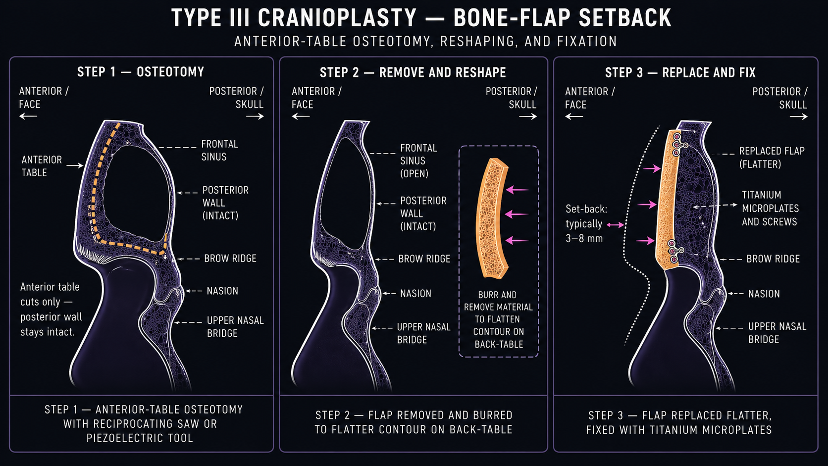
- Anterior table osteotomy. The bone wall over the frontal sinus is cut free as a single piece (or planned segments with VSP).
- Sinus mucosa preservation. The lining inside the sinus is left intact when possible. Both frontonasal duct patencies (the drainage paths from the sinus down into the nose) are verified.
- Reshaping. The freed bone flap is contoured externally; the surrounding bone (supraorbital rim, frontomalar transition, nasal bridge transition) is also burred to feminize the whole upper-face curve, not just the central bossing.
- Setback and fixation. The bone flap is replaced in a more recessed position and fixed with titanium or bioabsorbable plates and small screws. Some modern centers use titanium mesh as an adjunct when the bone is thin or focally defective.
- Closure. Soft tissue is repositioned, sometimes with a simultaneous brow lift, and the scalp is closed in layers. Drains may be placed for 24–48 hours.
Operating time varies widely with planning approach and surgeon experience — the VSP cadaveric study used 19 minutes for the setback itself, but the full FFS session including other procedures typically runs 6–10 hours.
Recovery
Specific timelines are less standardized than the technical descriptions. The numbers below come primarily from the 2014 Capitán 172-case series, which remains the most concrete published forehead-specific recovery data.
| Phase | What to expect |
|---|---|
| Day 0–2 | Hospital observation; head dressing; drains removed at 24–48h if used; significant edema (swelling) of the forehead and around the eyes |
| Day 7–8 | First in-person follow-up; dressing removed; sutures or staples removed depending on closure; bruising peaks around now |
| Weeks 2–6 | Most external swelling resolves; bruising fades; numb patches on forehead and scalp become apparent (and bothersome) |
| Weeks 8–12 | Hair starts growing through the beveled hairline scar if a pretrichial approach was used; the scar is at its most visible during this period before becoming concealed |
| Month 3 | Sensory recovery typically begins — the tingling/itching that signals nerve regeneration |
| Month 6 | Formal aesthetic review point; most subtle swelling resolved; cephalometric outcomes and patient satisfaction in published series stabilize around here |
| Month 6–12 | Final result; residual numbness in patches may persist permanently in a minority of patients |
Numbness is the most common medium-term complaint. The supraorbital nerves run right through the surgical field and are stretched, compressed, and (in patients with bony foramina) actively osteotomized free. Most patients have at least light-to-moderate paresthesia of the forehead and front of the scalp post-op; recovery begins around 3 months but can be patchy or incomplete.
The literature does not provide high-quality evidence on optimal compression, antibiotic duration, return-to-work timing, or activity restrictions specific to forehead surgery. Follow your surgeon's protocol; expect a wide range across surgeons.
Risks and complications
The pooled rate of complications across 10 systematic-review studies (673 patients) is 1.3%. That's low, but it understates a few things — specifically, the late risks that short follow-up misses.
| Complication | Pooled rate | Notes |
|---|---|---|
| Overall complication rate (Type III setback) | 1.3% (n=673) | From 10-study systematic review |
| Revision / reoperation | 0.4% | |
| CSF leak (cerebrospinal fluid) | 0.15% (1/673) | Most consequential acute complication |
| Anterior table mobility / pneumosinus dilatans | 0.45% | Fixation/healing failure |
| Transient frontal-branch facial nerve weakness | 0.15% pooled (likely undercounted) | Usually resolves in weeks |
| Infection / hardware extrusion | Rare; 0/43 in 2024 cohort | |
| Hematoma requiring drainage | Rare; 0/172 in one large series |
Sensory change (numbness, paresthesia) is reported as common but inconsistently quantified — most papers describe it qualitatively rather than giving a numeric rate. Plan on some, hope it resolves.
Alopecia and hypertrophic scarring are real risks at the incision site but rate data are weak across the literature.
Late sinus risks
The frontal sinus is opened in Type III surgery. That creates a small but genuine tail risk that doesn't show up in studies with short follow-up:
- Sinocutaneous fistula — a tract from sinus to skin. Reported case-by-case at 2–10 years post-op.
- Frontal mucopyocele / osteomyelitis — infected fluid collection or bone infection in or around the operated sinus. Rare case reports ~5 years out.
- Trace mucosal thickening on CT — found in 12.5% of patients (5/40) at mean 71 months follow-up in a 2026 imaging study. Importantly, none of these patients had clinically significant disease and zero needed sinus surgery within the first 6 years.
The reassuring read: clinically meaningful late sinus disease is uncommon in the first 5–6 years. The unreassuring read: nobody has high-quality 10+ year data, and the case reports show that fistula and mucopyocele can show up that late. Persistent forehead pain, tenderness, swelling, or drainage years post-op deserves prompt sinus imaging — don't write it off as unrelated to your old FFS.
What to ask in consult
| Question | Why it matters |
|---|---|
| What does my CT show — sinus depth, anterior table thickness, septations? | This determines which type of operation you'll have. If your surgeon can't show you on the imaging, that's a flag. |
| Type I, I+, II, or III? Why this one? | A surgeon who can't articulate the anatomical reason isn't doing the analysis. |
| Do you use virtual surgical planning or cutting guides? | Not mandatory, but adds safety on complex anatomy. |
| What's your fixation method — wires, plates, mesh? | Modern default is titanium or bioabsorbable plates/screws; mesh is an adjunct for weak or defective anterior table. |
| Coronal or pretrichial incision? Why this one for me? | Pretrichial only if you also need hairline advancement. |
| What's your revision rate, and what's covered? | Pooled revision rate is 0.4%; surgeons should know their own. |
| What's your protocol for late sinus complications? | Should have an answer; should not be "you'd just go to ENT." |
| How many Type III procedures have you done in the last year? | Volume matters for any cranioplasty. |
Gender-affirming framing
WPATH SOC8 (2022) and the international evidence-based consensus on facial gender surgery both classify FFS as medically necessary, not cosmetic. Hormone therapy is not a prerequisite. Patient-reported quality-of-life and gender-congruence improvements are well-documented in the multicenter prospective FFS literature.
The legal and insurance landscape is still inconsistent — many U.S. payers continue to deny FFS as cosmetic despite SOC8. Coverage is improving but uneven; some surgeons' offices have established appeals workflows. See the FFS overview for general cost and insurance context.
Sources
Foundational technique
- Ousterhout DK. (1987) — Feminization of the Forehead: Contour Changing to Improve Female Aesthetics (Plast Reconstr Surg) — original Type I/II/III taxonomy in feminizing cranioplasty.
- Capitán L et al. (2014) — Facial Feminization Surgery: The Forehead. Surgical Techniques and Analysis of Results (Plast Reconstr Surg) — 172-case series; the most concrete published recovery and operative-detail dataset (cephalometric setbacks, satisfaction 4.5/5, no CSF leaks, no dural injuries).
- Pansritum K et al. (2021) — Forehead and Hairline Surgery for Gender Affirmation (PRS Global Open) — 23-case combined forehead + hairline advancement series; grid-based frontal sinus border identification.
Operative series and technical advances
- Moghadam et al. (2024) — The Type I+ Forehead in Facial Feminization Surgery (Aesthetic Plast Surg) — 123-patient retrospective: 8 Type I+ vs 115 Type III; defines the hybrid technique for shallow-sinus forehead with thick anterior table.
- Gray et al. (2019) — Osseous Transformation with Facial Feminization Surgery: Improved Anatomical Accuracy with Virtual Planning (Plast Reconstr Surg) — cadaveric VSP vs no-plan: forehead setback time 19 vs 44 min, safety 100% vs 88%, volumetric accuracy 97% vs 79%.
Reviews and long-term follow-up
- Khetpal S et al. (2024) — Feminization of the Forehead: A Scoping Literature Review and Cohort Study of Transfeminine Patients (Aesthetic Plast Surg) — pooled complication rates: 1.3% overall, 0.15% CSF leak, 0.45% anterior-table mobility, 0.4% revision (≈673 patient literature pool).
- Jain-Poster et al. (2026) — Radiologic Evidence of Delayed Frontal Sinus Disease After Feminizing Frontal Cranioplasty (Otolaryngol Head Neck Surg) — 40 patients at mean 71 months; 12.5% trace mucosal thickening, no mucoceles or sinus surgeries within first 6 years.
Standards and frameworks
- Coon D et al. (2022) — Facial Gender Surgery: Systematic Review and Evidence-Based Consensus Guidelines from the International Facial Gender Symposium (Plast Reconstr Surg) — international consensus that facial gender surgery is medically necessary, not cosmetic.
- Coleman et al. (2022) — WPATH Standards of Care v8 — facial gender surgery medical necessity criteria; hormones not prerequisite.