Combining with FFS
Most patients seeking a feminizing transplant also want — or have already had — hairline advancement and forehead contouring. These three procedures share scalp territory and blood supply, and the literature is genuinely split on how they should be sequenced.
This page is scoped to the transplant-specific interactions with the rest of upper-face FFS: shared vasculature, scarred recipient beds, the simultaneous in-session protocol, the "transplant-first" rationale, and the practical realities of finding a surgical pair willing to coordinate. The basic candidacy split (advancement vs. transplant as primary) lives in Hairline Advancement; the broader sequencing framework across all of FFS lives in Procedure Staging. For recovery-specific interactions when grafting is delayed past FFS, see Recovery & Aftercare, and for whether to graft at all, see Candidacy & Timing.
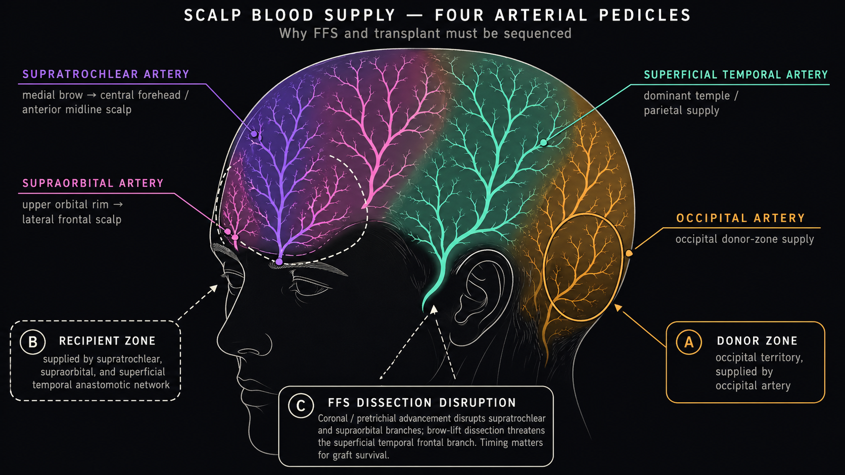
Why these procedures interact at all
The frontal scalp is supplied by three paired pedicle systems, all of which get touched by FFS:
- Supratrochlear and supraorbital arteries ascending from the medial brow into the central frontal scalp — directly in the field of forehead contouring and the lifted flap of any coronal/pretrichial advancement.
- Superficial temporal arteries entering laterally — the dominant supply to the frontotemporal transition zone where M-shape transplants are placed, and the territory most vulnerable to alopecia bands when temporal incisions cross a previously dissected scalp.
- Occipital arteries posteriorly — the donor zone for both FUE and FUT harvest. Largely spared by FFS but compressed by Mayfield/horseshoe positioning during long bone-work cases.
A hair graft is essentially an avascular cylinder for the first 48–72 hours (plasmatic imbibition) before neovascularization at roughly 1 mm/day from the recipient bed. Anything that reduces recipient-bed perfusion reduces graft take. This is the central mechanism behind every staging argument below.
The dissection planes also overlap. Pretrichial and coronal exposures use the subgaleal plane (loose areolar tissue between galea aponeurotica and pericranium) — the same plane in which transplant recipient sites are made. Type III forehead contouring goes a level deeper to subperiosteal for the osteotomy. After healing, both planes scar in ways that change the angle and depth at which a transplant blade can safely make slits without transecting deeper structures or the previously elevated flap's perforators.
The literature is split three ways
Three defensible sequences exist in the published record. Each camp has a coherent argument and a different idea of what the most important risk is.
| Sequence | Core argument | Weakness |
|---|---|---|
| Simultaneous (transplant during FFS) | One anesthetic, one recovery, one trip. FUT-from-coronal-strip uses tissue that would otherwise be discarded. | Recipient bed is freshly dissected and edematous; no FFS-specific counted-graft survival data. |
| Staged FFS-first (transplant ≥6–12 mo after) | Recipient bed is fully revascularized; pretrichial scar position and final relapse are visible; transplant surgeon can be a dedicated hair-restoration team. | Two recoveries, two travel windows, and patients with M-shape-dominant dysphoria live with a partial result for a year. |
| Transplant-first (FFS ≥9–12 mo after) | Advancement does not fix lateral recession. Building the M-shape correction first gives the advancement a feminized hairline shape to lift. | Subsequent pretrichial incision passes through transplanted tissue and creates a small zone of shock-loss alopecia. |
There is no head-to-head graft survival study comparing these in the FFS context with counted-hair-per-cm² methodology at one year. Until one exists, the choice rests on patient anatomy, surgeon philosophy, and logistics rather than on data.
The simultaneous protocol
The reference paper is Capitán, Simon, Kaye, and Tenorio (2017) — Facial Feminization Surgery: Simultaneous Hair Transplant during Forehead Reconstruction (Plastic and Reconstructive Surgery 139(3):573–584). Sixty-five transfeminine patients; FUT-from-coronal-strip; no vascular complications reported.
The protocol uses FUT-from-coronal-strip: the strip removed during pretrichial/coronal access is dissected on the back table into individual follicular units while bone work proceeds. The intraoperative sequence is bone work → forehead set-back → scalp advancement → closure → recipient site creation in the newly advanced scalp → graft placement. Recipient zones are the frontotemporal corners and any residual M-shape after advancement is closed. Graft counts are typically several hundred to ~1,000 — well below the 2,000–3,500 of a dedicated session. Follow-up was up to 12 months by clinical photography only.
The data gap behind the simultaneous protocol
Hair-restoration literature reports survival as 80–95% take in unscarred recipient beds, counted graft-by-graft. Capitán 2017 reports survival qualitatively. No FFS-specific study has counted grafts in 1 cm² grids at one year to give a comparable number for simultaneous-with-advancement transplants. Whether take drops 5%, 20%, or more in this context is unmeasured. This is the single most important data gap in the simultaneous-vs-staged debate.
The biological argument against simultaneous grafting is plasmatic imbibition in edematous tissue: a 6–10 hour FFS produces real frontal edema by case end. The Capitán protocol works around this by grafting late in the case into the advanced scalp (which is less manipulated than the excised strip's territory) and limiting graft counts. The pragmatic constraint is that simultaneous grafts cannot be re-spaced if the advancement settles asymmetrically over the next 6–12 months — the design is committed to the immediate intraoperative geometry.
Other groups practicing simultaneous FUT-from-strip during FFS include the Deschamps-Braly San Francisco group (anecdotal) and chapter-form descriptions from the Yale Alperovich group. The Plastic Surgery Key FFS chapter, referencing a 492-patient transfeminine cohort, treats simultaneous transplant as one valid pathway rather than the default.
The staged approach (transplant ≥6 months after FFS)
The mainstream hair-restoration position. The structure: FFS at month 0 → healing and scar maturation through months 6–12 → transplant session at month 9–12 into healed tissue → optional second transplant 9–12 months later.
The 6–12 month interval is the standard recommendation but is not derived from FFS-specific data. It comes from general hair-restoration literature on grafting into post-traumatic and post-surgical scars (Unger, Shapiro, Unger, and Unger (2010) — Hair Transplantation, 5th ed., the foundational reference). Twelve months is the more commonly recommended figure when scalp advancement was substantial, because remodeling continues past month 6 and final scar width is not stable until ~12 months.
The case for staging:
- Recipient bed is fully revascularized; perforator network has remodeled around the new scar.
- The design canvas is final. The pretrichial scar's exact position, the settled relapse (1.98–2.51 mm of forehead lengthening at 2 years per Lee et al. 2021), and post-edema temple width are all visible.
- Grafts can be placed across the scar to camouflage it (see below) once the scar is mature.
- The transplant surgeon can be a dedicated hair-restoration specialist, not whoever the FFS surgeon's in-house grafter is.
The cost: two recoveries, two travel windows for patients flying to FFS centers, and a 9–12 month gap living with a partial result if the M-shape is the patient's primary dysphoria trigger.
The "transplant-first" approach
This is the option that gets least airtime in consult brochures and is specifically called out in the Plastic Surgery Key FFS chapter referencing a 492-patient transfeminine cohort. The argument rests on a single anatomical fact: a 1.8–2.2 cm pretrichial advancement lowers the central hairline but pulls the M-shape forward in parallel — the corners are still M-shaped, just at a lower height. In Khetpal et al. (2024), 79% of transfeminine FFS patients had lateral recession (Aesthetic Plast Surg).
For a patient whose dominant dysphoria trigger is the M-shape rather than central height, advancement is the wrong primary procedure. Transplant builds feminine temple corners that advancement cannot. Doing transplant first, then advancement 9–12 months later, gives the surgeon a feminized hairline shape to advance — the advancement preserves the shape rather than having to create it.
The vascular argument runs the other direction here: transplanting into virgin scalp is the lowest-risk grafting environment, so doing it first protects graft take. The cost is that the subsequent advancement passes a pretrichial incision through transplanted tissue, which does transect some recently established follicles at the leading edge and creates a small zone of shock-loss alopecia adjacent to the scar. This is a real tradeoff with no published outcome data quantifying which order preserves more total surviving feminine hair.
The Plastic Surgery Key chapter recommends transplant-first selectively — specifically for patients whose M-shape is dominant, donor density is good, and androgenetic alopecia is medically stable. It is not a universal recommendation.
What transplants can do with the pretrichial scar
The scar from advancement is the most concrete constraint on a future transplant. Two questions matter.
Across the scar? Yes, with caveats. The trichophytic / hyperbeveled incision design (Ramirez, Ende, and Kabaker 2009; Arch Facial Plast Surg) is engineered so native hair grows through the scar to camouflage it. After scar maturation (≥12 months), single-hair FUE grafts placed at acute angles into and posterior to the scar are the standard camouflage technique. Take rates are typically reported as 10–30% lower than in unscarred recipient sites, which is why transplant surgeons over-graft scar zones by a corresponding margin.
Anterior to the scar — creating a new lower hairline below the advanced one? The skin anterior to a pretrichial scar is original forehead skin with its own mature blood supply, but a transplant there cannot grow a feminine transition without bridging the scar with single-hair grafts at multiple densities. If more lowering is needed, a second advancement or a tissue expander is more predictable. The scar-camouflage indication dominates.
A coronal scar (posterior to the hairline, in hair-bearing scalp) is much more forgiving — grafts placed anterior to it operate in relatively normal tissue with intact frontal perforators. This is one quiet argument for coronal access in patients who anticipate eventual transplant. The countervailing argument — that a coronal incision in someone who actually needed pretrichial lowering is a hard mistake to fix — remains the dominant published view.
Vascular concerns when re-operating on a previously dissected scalp
This is the scenario where the literature is most cautious and the fewest published numbers exist.
A coronal/pretrichial flap raises the entire frontal scalp on its lateral and posterior pedicles, cutting the supratrochlear and supraorbital perforators that ascend from the brow. Those perforators do reconnect over months as the flap revascularizes from the deeper plane and from the lateral scalp, but the density of small perforators in the central frontal scalp is reduced long-term.
Re-operating in this reduced-perforator tissue — a second pretrichial design, a brow revision, or aggressive temporal recipient-site creation — increases the risk of alopecia bands between scars: strips of relative ischemia where neither the new nor the old blood supply reaches reliably.
Revision cases are different
The Ahn 2019 multiplane series (PRS) explicitly excluded patients with prior scalp surgery from the cohort. Modern revision-focused FFS literature warns that a second pretrichial design has higher alopecia risk between scars because of compromised vascularity. The reassuring complication rates in primary FFS series do not transfer to revision cases.
This is the strongest argument for the standard 9–12 month healing interval between advancement and a subsequent transplant: it gives the dissected scalp time to re-perforate before further insult.
The transplant interaction with rhinoplasty is purely additive operative time, not vascular. Forehead-then-rhinoplasty is the consensus sequence (Bellinga et al. 2017); transplant slots in at the end of the upper-face block, after closure, before the team transitions to the nose. None of the major bundled-FFS series isolates simultaneous-transplant graft survival.
Decision framework
Right sequence depends on five inputs. Use this in consult — and use it as a filter on the recommendation you're given.
| Input | Pushes toward simultaneous | Pushes toward staged (FFS first) | Pushes toward transplant first |
|---|---|---|---|
| Dominant problem is central height | Yes (advancement does the work) | Yes (advancement does the work) | No |
| Dominant problem is M-shape / temples | No (advancement won't fix it) | No (still need transplant after) | Yes |
| Donor density is good, AGA is stable | Permissive | Permissive | Required |
| Active androgenetic alopecia | Defer transplant entirely until stable | Defer transplant entirely until stable | Contraindicated |
| Patient travels for FFS, can't return | Strong push | Hard logistics | Hard logistics |
| Patient values predictable graft take | Weaker case (no objective survival data) | Strong case (virgin/healed bed) | Strong case (virgin bed) |
| Type III forehead with major setback | Permissive (Capitán protocol designed for this) | Permissive | Caution: subsequent advancement scar bisects new transplant |
| Smoker / poor wound healing | Avoid bundling | Avoid bundling | Stage everything; address smoking first |
| Revision case (prior FFS scalp surgery) | High risk; most surgeons decline | Mandatory | Often best (transplant into less-disturbed lateral scalp) |
The single highest-yield clarifier in consult is asking the surgeon to name your dominant feminization deficit: central height, M-shape, density, or shape irregularity. Each maps to a different primary procedure, and once the primary is named, the others slot in around it.
The surgeon-availability problem
This is the practical reality skipped in literature reviews. Almost no surgeon does both high-volume FFS and high-volume hair restoration. The short overlap roster as of recent years:
- FACIALTEAM (Marbella) — Capitán, Simon, Bellinga, plus a dedicated in-house transplant team. The Capitán 2017 protocol means the same trip can include simultaneous transplant; staged transplant on returning patients is also offered.
- Deschamps-Braly (San Francisco) — performs FFS with transplant collaboration; in-session FUT during forehead work is offered.
- Spiegel (Boston) — FFS-focused; collaborates with hair-restoration surgeons rather than offering in-house simultaneous transplant.
- Most other major FFS surgeons (Plastic Surgery Institute, Crane, Mardirossian, Mommaerts, Telang, etc.) refer transplant out, generally to a hair-restoration practice in the same metro.
For most patients this means two surgeons in two cities, and coordination is the patient's job. Practical issues: the transplant surgeon can't see the FFS plan until the FFS surgeon shares it (and FFS surgeons rarely write hair-restoration-friendly operative plans); pretrichial scar position usually constrains transplant design and needs to be seen at ≥6 months; insurance treats the procedures separately even when bundled medically; and travel timing compounds when the two surgeons are in different countries.
The published FFS literature does not address coordination explicitly, but it shapes which sequence is realistic at least as much as the anatomy.
Caveats and data gaps
- Capitán 2017's 65-patient survival data are qualitative; no formal trichoscopy or counted-graft follow-up is published.
- The 492-patient cohort cited via Plastic Surgery Key is not an open-access outcomes paper; the chapter summarizes a single institution's experience.
- "Alopecia bands between scars" is a clinically real entity but is mostly reported in revision case series and revision-focused chapters rather than prospective cohorts.
- Smoking, prior scalp surgery, and revision status are exclusion criteria in most major FFS series — the reassuring complication numbers do not apply to patients with the most complicated transplant decisions.
- The 9–12 month interval recommendation is widely repeated but is not derived from FFS-specific study; it is a graft-physiology and scar-maturation default.
Sources
- Capitán L, Simon D, Kaye K, Tenorio T (2017) — Facial Feminization Surgery: Simultaneous Hair Transplant during Forehead Reconstruction (Plastic and Reconstructive Surgery 139(3):573–584). 65-patient single-stage protocol; FUT-from-coronal-strip; qualitative survival reporting.
- Plastic Surgery Key — Facial Feminization Surgery and Facial Gender Confirmation Surgery — chapter referencing the 492-patient transfeminine cohort; explicit transplant-first recommendation for M-shape-dominant cases with stable AGA.
- Khetpal S, et al. (2024) — Feminization of the Forehead: A Scoping Literature Review and Cohort Study of Transfeminine Patients (Aesthetic Plastic Surgery). 79% lateral recession prevalence — central evidence for the transplant-first M-shape argument.
- Lee K, et al. (2021) — Forehead Reduction Surgery via an Anterior Hairline Pretrichial Incision in Asians: A Review of 641 Cases (Aesthetic Plast Surg). 1.98–2.51 mm relapse at 2 years informs the staged-transplant design canvas argument.
- Ramirez AL, Ende KH, Kabaker SS (2009) — Correction of the High Female Hairline (Archives of Facial Plastic Surgery). Trichophytic / hyperbeveled incision design that permits later transplant grafting through the mature scar.
- Unger W, Shapiro R, Unger R, Unger M (2010) — Hair Transplantation, 5th ed. (Informa Healthcare). Standard reference for grafting into post-surgical scars; basis for the 6–12 month interval default.
- Bellinga RJ, Capitán L, Simon D, Tenorio T (2017) — Rhinoplasty in Facial Feminization Surgery: 200 Consecutive Cases (Facial Plastic Surgery). Forehead-then-rhinoplasty bundle context.
- Kim S, et al. (2024) — Perioperative Estrogen Hormonal Therapy Does Not Increase Venous Thromboembolism Risk in Facial Feminization Surgery (PRS). 953-patient Deschamps-Braly cohort; 0.10% VTE; bundled-FFS safety baseline.
- Coleman E, et al. (2022) — WPATH Standards of Care v8 — hairline / forehead surgery as medically necessary; relevant to insurance coordination across staged FFS + transplant pathways.